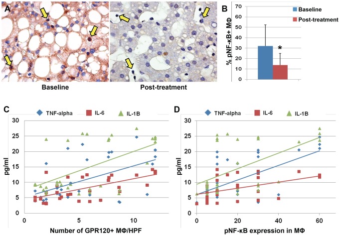
Figure 4. Nuclear expression of phosphorylated (p) NF-κB by Macrophages in pediatric NAFLD patients.
A) Immunohistochemistry for serine-311-phosphorylated NF-κB (pNF-κB) in pediatric NAFLD biopsies before and after DHA treatment. In parallel with GPR120 expression, the DHA treatment determined a reduction of pNF-κB expression and its nuclear translocation in macrophages (A, yellow arrows). Original magnification: 40×. B) Histogram shows the significant reduction of pNF-κB nuclear translocation in macrophages after DHA treatment. Data are shown as Means ± Standard Deviation. * = p<0.05. C–D) The number of GPR120-positive macrophages (C) and the pNF-κB nuclear expression in macrophages. (D) were correlated with serum level of inflammatory cytokines.

