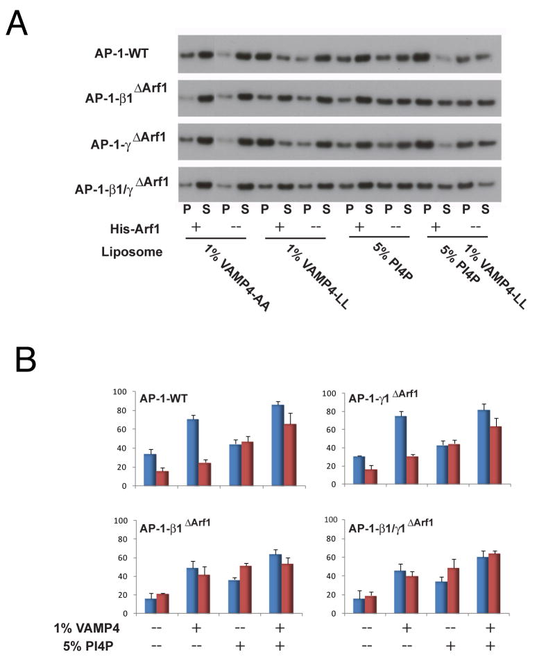
Figure 7. Reconstitution of membrane recruitment and activation by Arf1 and cargo.
(A) Recruitment of the AP-1 core to peptidoliposomes by lipid sedimentation assay. Liposomes were made of 5% DOGS-NTA:POPC:POPE, 1% VAMP4-LL/AA lipopeptide, 1% VAMP4 (1-51) lipopeptide, 5% PI(4)P, or both PI(4)P and VAMP4 lipopeptide. AP-1 cores (20 nM) were incubated with or without His-Arf1-GTP (50 nM) ultracentrifuged to separate the pellet (P) and supernatant fractions (S). Fractions were immunoblotted with anti-μ1 (A), and quantified using ImageJ. The AP-1 membrane binding percentage was calculated according to the formula (P/P+S) × 100% (B) Quantification of sedimentation data. Assays containing Arf1 are colored code in blue and without Arf1 in red. The error bars represent the standard deviation of three replicates.

