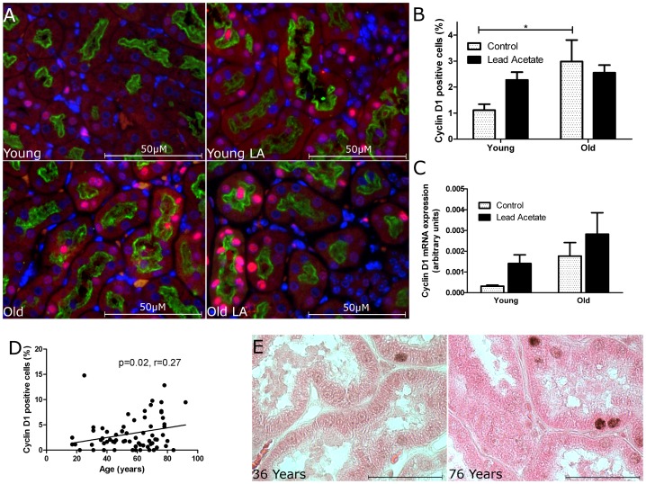
Figure 3. Baseline expression of cell cycle protein Cyclin D1 is higher in tubular cells of old kidneys than tubular cells of young kidneys.
(A) Representative photographs of Cyclin D1 (red) immunostaining of kidney sections from young and old mice with or without lead acetate treatment. LTL (green) stains the brush border membrane of the proximal tubule; original magnification 400×. (B) Quantification of tubular cells with Cyclin D1 positive nuclei. (C) Analysis of Cyclin D1 mRNA expression. (D) Quantification of Cyclin D1 positive cells in renal transplant implantation biopsies (n = 36) and healthy renal tissue from nephrectomised patients (n = 22) shows a significant positive correlation between tubular Cyclin D1 expression and chronological age. (E) Representative photographs of Cyclin D1 immunostaining of kidney sections from a younger (36 years) and an older (76 years) human kidney; original magnification 400×. n = 5 for mice. Data are mean values ± SEM. *P<0.05.

