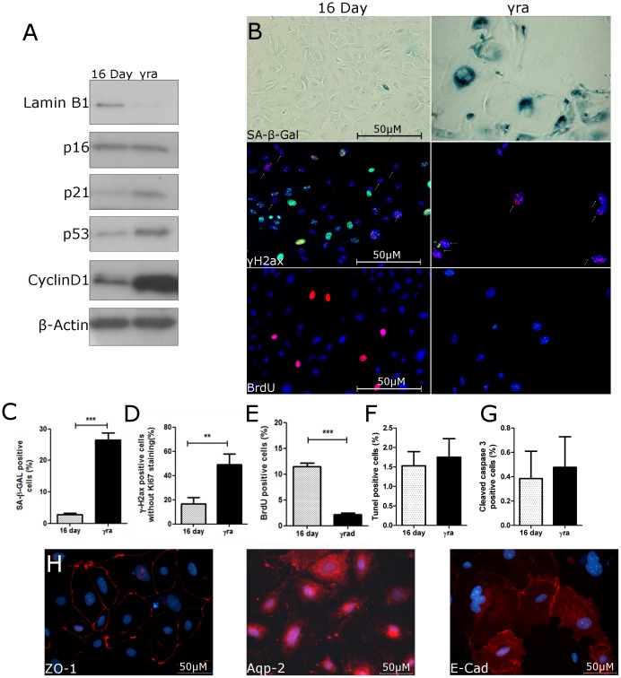
Figure 6. γ-irradiation induces senescence in PTEC and leads to increased Cyclin D1 expression.
PTEC were isolated and grown for 6 days in culture before being exposed to 10 Gy γ-irradiation. After y-irradiation, cells were split and grown for 10 days and tested for senescence markers. Controls were grown for 6 days, split and grown for another 10 days. (A) Representative immunoblots for Lamin B1 and cell cycle regulators p16INK4a, p21, p53, and Cyclin D1. (B) Representative photographs of SA-β-Gal, γH2AX and BrdU; original magnification 400×. Quantification of (C) SA-β-Gal, (D) γH2AX+/Ki-67−, (E) BrdU, (F) TUNEL, and (G) cleaved caspase 3 positive cells in cultures of control and γ-irradiated PTEC. (H) Representative photographs of epithelial markers ZO-1, Aqp-2, and E-Cadherin in γ-irradiated PTEC; original magnification 400×. Data are mean values ± SEM. **P<0.01; ***P<0.001.

