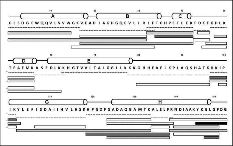
Figure 3.

Digest map of apoMb labeled with 10 mM pLeu. Labeled apoMb was digested with a combination of trypsin and chymotrypsin. White bars represent unlabeled peptides, while labeled peptides are shown in gray (light gray bars carry one label; dark gray bars carry two labels and the black bar carries four labels). Dashed lines represent native peptides. Helical secondary structure is represented by cylinders labeled A-E, G and H. Helix F of holomyoglobin (H82-H97) is disordered in native apoMb at neutral pH (see ref. 27).
