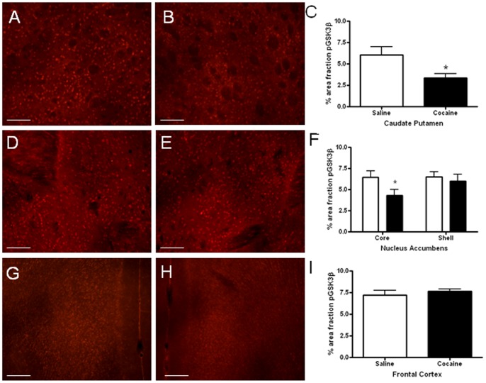
Figure 3. Immunofluorescence labeling of pGSK3β in the mouse brain.
Photomicrographs of pGSK3β immunolabeling in the caudate putamen of a mouse injected with saline (3A) or cocaine (3B). Acute administration of cocaine reduced the phosphorylation of GSK3β compared to saline administration in the caudate putamen (*p<0.05; 3C). Photomicrographs of pGSK3β immunolabeling in the nucleus accumbens of mice injected with saline (3D) or cocaine (3E). Levels of phosphorylated GSK3β were significantly lower in the core of the nucleus accumbens (*p<0.05), but not the shell (p>0.05), following cocaine administration (3F). pGSK3β immunolabeling in the frontal cortex of mice injected with saline (3G) or cocaine (3H). No differences were found in pGSK3β immunolabeling in the frontal cortex (3I). Mean ± SEM, (N = 9–10 mice/group). * p<0.05. Scale bar = 50 µm.

