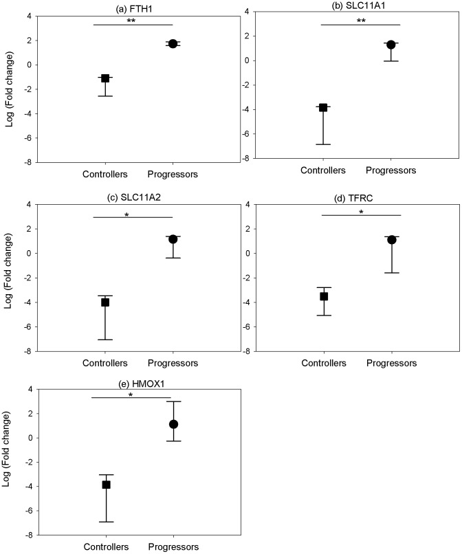
Figure 3. Expression of iron regulatory genes in PBMCs isolated from unvaccinated controllers and progressors at post-mortem.
Gene expression of a) ferritin heavy chain b) solute carrier family 11 member A1, and c) solute carrier family 11 member A2, d) transferrin receptor 1 and e) heme oxygenase 1 in unstimulated PBMCs isolated at post-mortem. The fold change from naïve baseline levels was determined using RT-PCR. To determine statistically significant differences of relative gene expression, a two-sample t-test was performed where * and ** represent P-values of <0.05 and <0.01, respectively. The data were from 3 animals. The symbol represents the median and the error bars represent the range.

