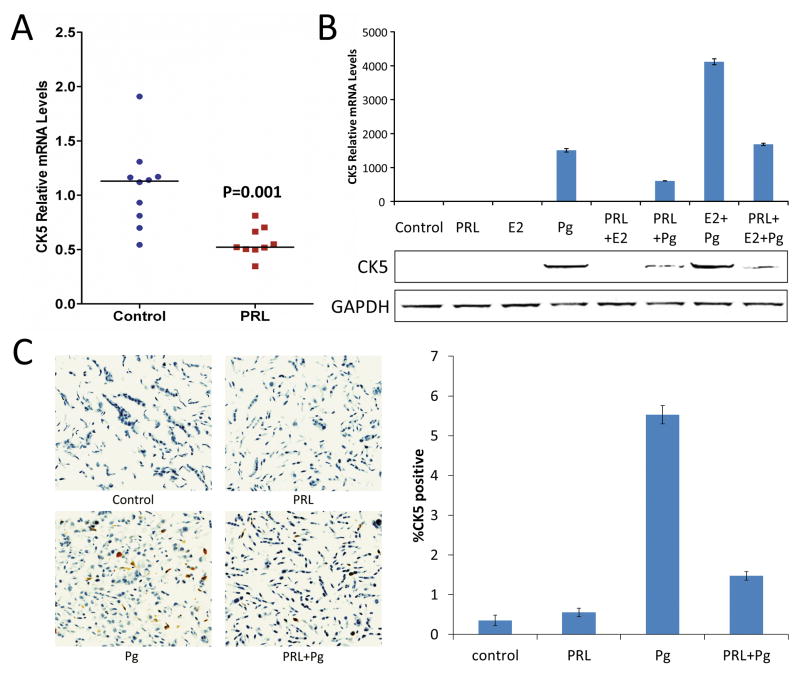
Figure 1. Prolactin suppresses CK5 mRNA and protein levels in human breast cancer cells.
(A) qRT-PCR analysis of CK5 mRNA extracted from T47D xenograft tumors in mice treated with either vehicle or prolactin for 48 h. Individual values are plotted and horizontal markers indicate median levels (P=0.001 by Mann-Whitney U test). (B) qRT-PCR (top) and immunoblot (bottom) analysis of CK5 mRNA and protein levels, respectively, in extracts of cultured T47D treated with vehicle (Control), Prolactin (PRL), β-Estradiol (E2), or R5020 (Pg) for 48 h. (C) Immunocytochemistry using DAB chromogen (brown) for CK5 and hematoxylin counterstain of sections of formalin-fixed, paraffin-embedded pellets of T47D cells treated with hormones as indicated for 24 h. Representative images are shown (top) with quantification for cellular CK5-positivity plotted (bottom). In Pg-treated cells, the percentage of CK5-positive cells was 3.8 times higher (95% CI: 1.6, 8.8, P=0.005) than in prolactin+Pg-treated cells.

