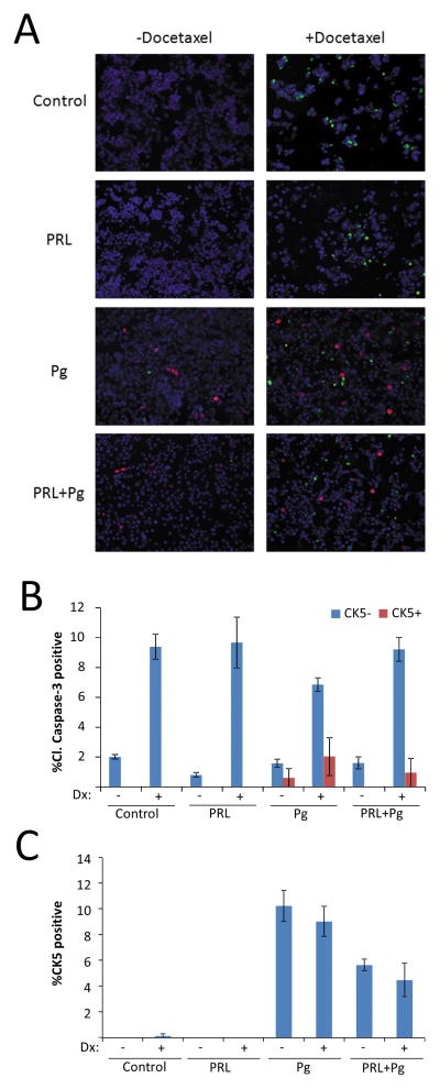
Figure 2. Prolactin suppresses the fraction of chemoresistant Pg-induced CK5-positive cells.
(A) Representative images of multiplexed immunocytochemistry of T47D cell cultures treated with vehicle (Control), Prolactin (PRL), β-Estradiol (E2), or R5020 (Pg) for 24 h before addition of docetaxel (Dx) or vehicle for 48 h and stained for CK5 (red), cleaved caspase-3 (green), and DAPI (blue). (B) Bar graph of percent of CK5-negative (blue bars) or CK5-positive (red bars) cells that were positive for cleaved caspase-3. Quantification revealed a four-fold reduced rate of cleaved caspase-3 staining in CK5-positive cells than in CK5-negative cells (odds ratio 0.25, 95% CI: 0.11, 0.56, P<0.001) (C) Overall percentage of cells positive for CK5 in the cultures shown in panel A. Odds of CK5 positivity were 50% lower for cells treated with prolactin plus Pg (5.7% CK5+) than for cells exposed to Pg alone (10.3% CK5+) (odds ratio= 0.50, 95% CI: 0.39, 0.64, P<0.001).

