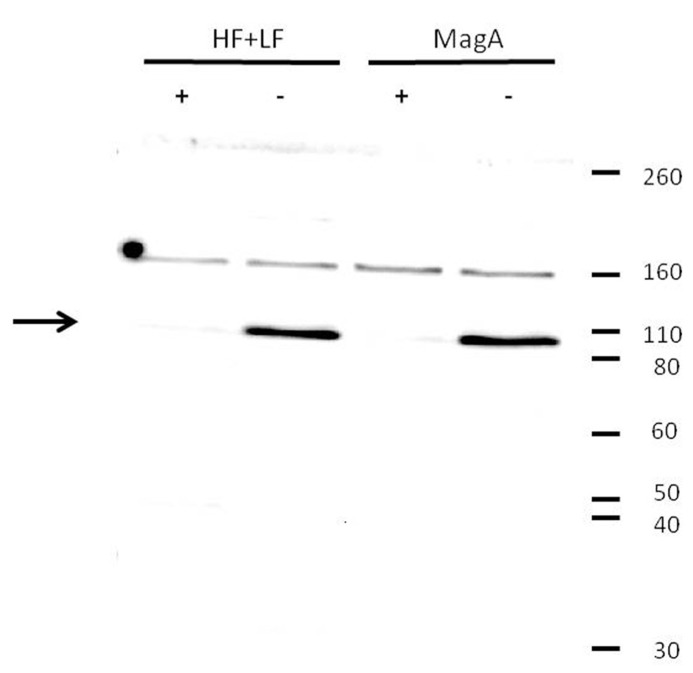
FIGURE 4.
Decrease in transferrin receptor expression upon iron supplementation. Tumor cells were cultured in the presence (+) or absence (-) of iron supplementation, lysed, and analyzed by Western blot using a primary antibody to transferrin receptor. Both MagA- and HF + LF-expressing cells showed greater immunoreactivity toward the soluble form of transferrin receptor (arrow) in cells cultured in the absence of iron supplementation. Under reducing conditions, soluble transferrin receptor migrates at a M.W. of approximately 95K. Protein M.W. standards are indicated on the right margin.

