
Figure 1.
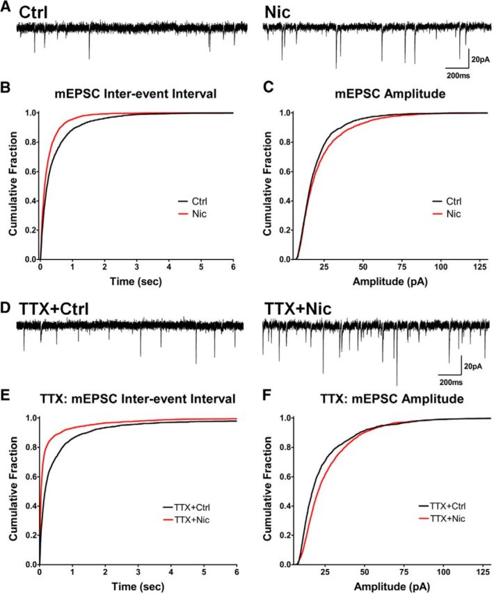
Nicotine treatment increases mEPSC frequency and amplitude generated by native AMPARs independently of AP-driven activity. A, Representative traces of mEPSCs in cells treated with nicotine for 1–3 h (Nic) versus controls (Ctrl). B, Cumulative histograms of mEPSC interevent intervals showing that the nicotine treatment increased the frequency of events (decreased the interevent intervals; Ctrl vs Nic, n = 2800 events for both, p < 0.0001, KS). C, Amplitude histograms showing that the nicotine treatment increases mEPSC amplitude (Ctrl vs Nic, n = 2800 events for both, p < 0.0001, KS). D, Representative traces of mEPSCs in cells treated with TTX to block APs while incubated with nicotine or control solution. E, Reduced interevent intervals, meaning increased mEPSC frequency, in cells treated with nicotine + TTX for 1–3 h (Ctrl + TTX vs Nic + TTX, n = 2500, 2600 events, p < 0.0001, KS). F, TTX did not prevent nicotine from increasing mEPSC amplitude (Ctrl + TTX vs Nic + TTX, n = 2400, 2600 events, p < 0.0001, KS).