Figure 2.
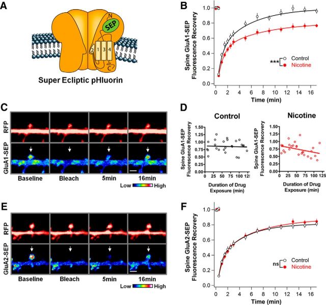
Nicotine stabilizes GluA1s on dendritic spines in hippocampal cell culture. A, Depiction of an AMPAR containing a transgenic GluA subunit tagged with SEP, a pH-sensitive GFP that only fluoresces while on the cell surface. B, GluA1-SEP fluorescence on spines of neurons treated with 1 μm nicotine for 1–3 h in cell culture recovers less in 16.5 min after photobleaching than do corresponding controls, indicating decreased mobility (Ctrl vs Nic: 96 ± 3 vs 77 ± 2%, n = 24,24; p = 0.00002, WC). C, Control condition images of GluA1-SEP in dissociated culture with accompanying cytosolic-RFP images. Top and bottom arrows indicate bleached spines. Scale bar, 2 μm. D, The nicotine effect on GluA1-SEP FRAP occurs gradually over 2 h. Cultures were transferred into a perfusion chamber with 1 μm nicotine and imaged for 2 h. Data points represent the final recovery point from different FRAP experiments. Values were fit with a linear regression demonstrating a trend in nicotine-treated cultures, but in not control cultures (control: slope −0.03 ± 0.12%, y-intercept 87.18 ± 8.42%, n = 25, r2 = 0.0026, p = 0.8087; nicotine: slope −0.22 ± 0.10%, y-intercept 85.85 ± 6.86%, n = 27, r2 = 0.1429, p = 0.0519). E, Control condition images of GluA2-SEP FRAP in dissociated culture with accompanying cytosolic-RFP images. F, Nicotine has no effect on GluA2-SEP mobility, yielding a recovery equivalent to control (Ctrl vs Nic: 81 ± 3 vs 84 ± 3%; n = 15,17; p = 0.49).

