Figure 1.
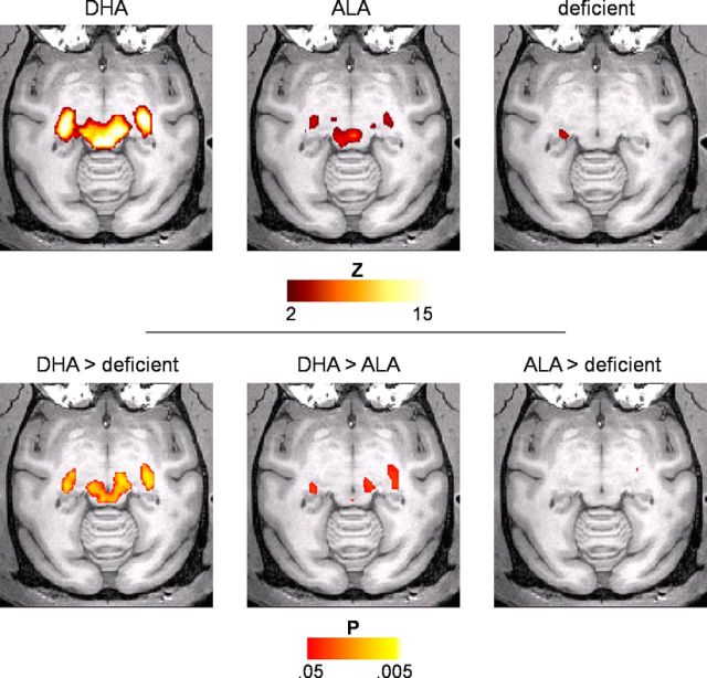
Connectivity with primary visual cortex in the LGN and SC is reduced in animals without dietary DHA. Top, Connectivity maps (|Z| > 2) within thalamus and superior colliculus show connectivity strength from V1 to LGN and SC in the order of DHA > ALA > deficient. Bottom, Pairwise group comparisons revealed significant (p < 0.05, Wilcoxon exact, two-tailed) differences between the DHA-fed and deficient groups (left) and between the DHA and ALA groups (middle). No significant differences were observed between the ALA-fed and deficient groups (right).

