Figure 3.
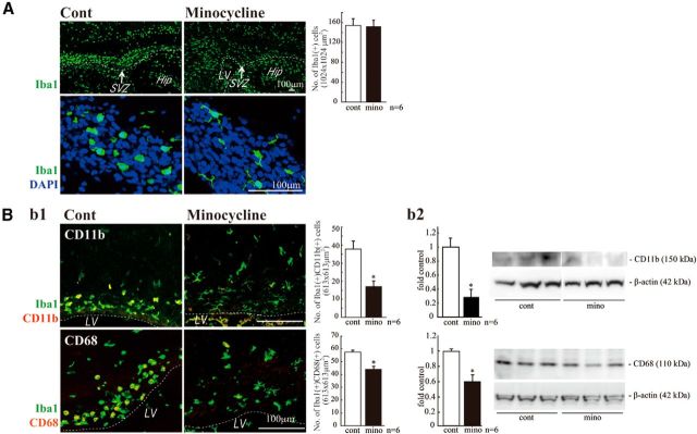
Minocycline suppressed microglial activation in vivo. A, Effects of minocycline on the number of Iba1+ cells in the SVZ and their morphologies. Minocycline was administered by intraperitoneal injection for 3 d beginning at P2 (30 mg/kg/d, P2-P4, n = 6/group). Sagittal sections of minocycline-treated forebrains were immunostained for Iba1 (green) followed by DAPI staining (cyan). Although the number of Iba1+ microglia in the SVZ did not change (graph), their shape shifted from an amoeboid type to a more ramified type by minocycline (bottom). Bb1, Effects of minocycline on the expression of activation markers and the morphologies of microglia. Sagittal sections of minocycline-treated forebrains were immunostained for Iba1 (green), and CD11b (red), and CD68 (red). Minocycline significantly decreased the number of cells positive for CD11b or CD68. The morphologies of the cells were also changed from amoeboid shape to more ramified shape. Bb2, The significant decrease in the expression of CD11b and CD68 was confirmed by Western blotting of the SVZ as well. *p < 0.05 (Student's t test). Data are mean ± SEM. Similar results were obtained in three independent experiments.

