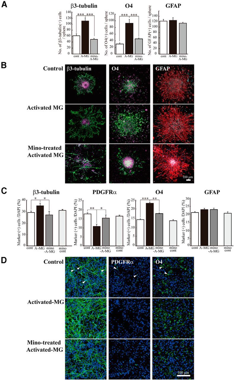
Figure 6.

The reproduction of the enhancement of neurogenesis and oligodendrogenesis by activated microglia in vitro. Microglia cultured independently of neurosphere on transwells were activated by LPS (10 ng/ml, 30 min) in the presence or absence of minocycline (10 μm), washed carefully, and the transwells were set onto the neurospheres or dissociated cells from neurosphere in prodifferentiation conditions. After differentiation periods suitable for neurons (7 d) or oligodendrocytes (11 d), neurospheres were stained for β3-tubulin (green), PDGFRα (green), O4 (green), GFAP (red), and TOTO3 (cyan). To check the effects of minocycline alone, dissociated cells were incubated in the presence of minocycline (10 μm) for 7 d. A, Quantification of the numbers of neurons, oligodendrocyte progenitors, or astrocytes differentiated from neurospheres cocultured with activated microglia in the presence or absence of minocycline. ***p < 0.001 (Tukey's test by ANOVA). n = 12 neurospheres/group. Data are mean ± SEM. B, Representative immunostained images of neurospheres cocultured with activated microglia in the presence or absence of minocycline. C, The effects of activated microglia on differentiation of single cells dissociated from neurospheres in the presence or absence of minocycline. The effects of minocycline alone were also shown (mino-cont in each graph). *p < 0.05, **p < 0.01, ***p < 0.001. (Tukey's test by ANOVA). n = 12 neurospheres/group. Data are mean ± SEM. D, Images of cells immunostained for differentiation markers. Arrowheads indicate the representative cells positive for the differentiation markers.
