
Figure 4.
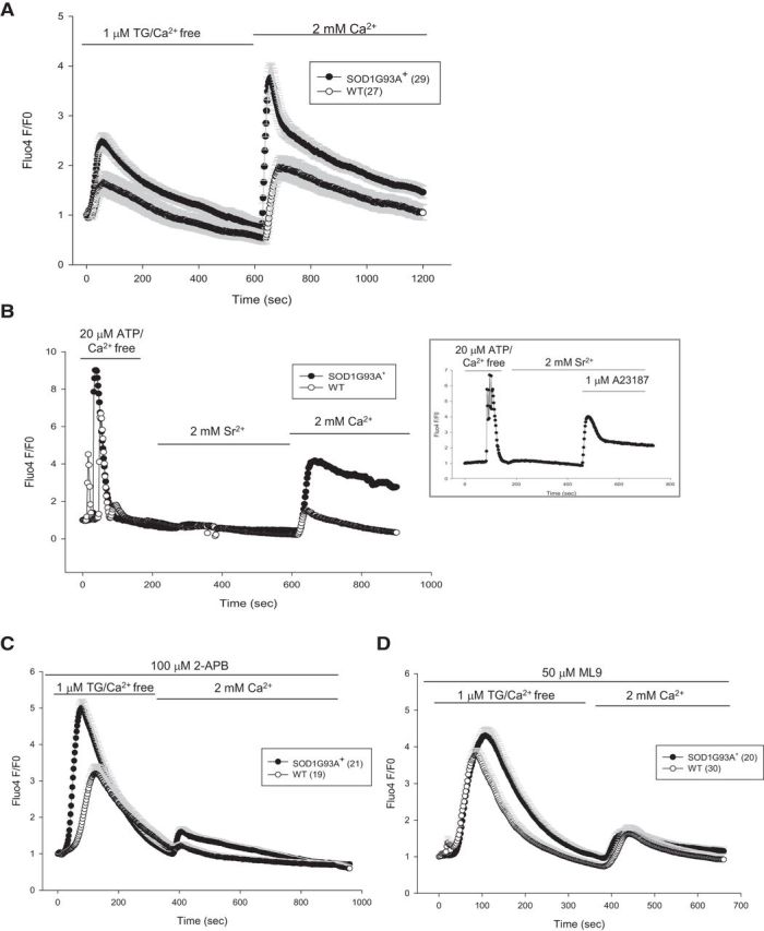
ER calcium-independent, abnormal SOCE in SOD1G93A+ astrocytes. A, SOCE in astrocytes was induced by ER calcium depletion with 1 μm TG in calcium-free medium, followed by perfusion with 2 mm calcium containing medium. The peak of SOCE and rate of entry are higher in SOD1G93A+ astrocytes. B, SOCE induced by 20 μm ATP/calcium-free medium to deplete ER calcium, but instead of 2 mm calcium, 2 mm strontium was used to discriminate between Orai and TRPC channels. At the end of the experiment, 2 mm calcium was perfused. The graph in the inset shows the same experiment, but 1 μm calcium ionophore A23187 was perfused at the end to show that Fluo4 fluorescence responds to strontium. C, Average traces of SOCE in astrocytes pretreated with the SOCE inhibitor 2-APB (100 μm) for 10 min. D, Cells were pretreated with another SOCE inhibitor, ML-9 (50 μm, 10 min), before imaging. All error bars are SEM.