
Figure 5.
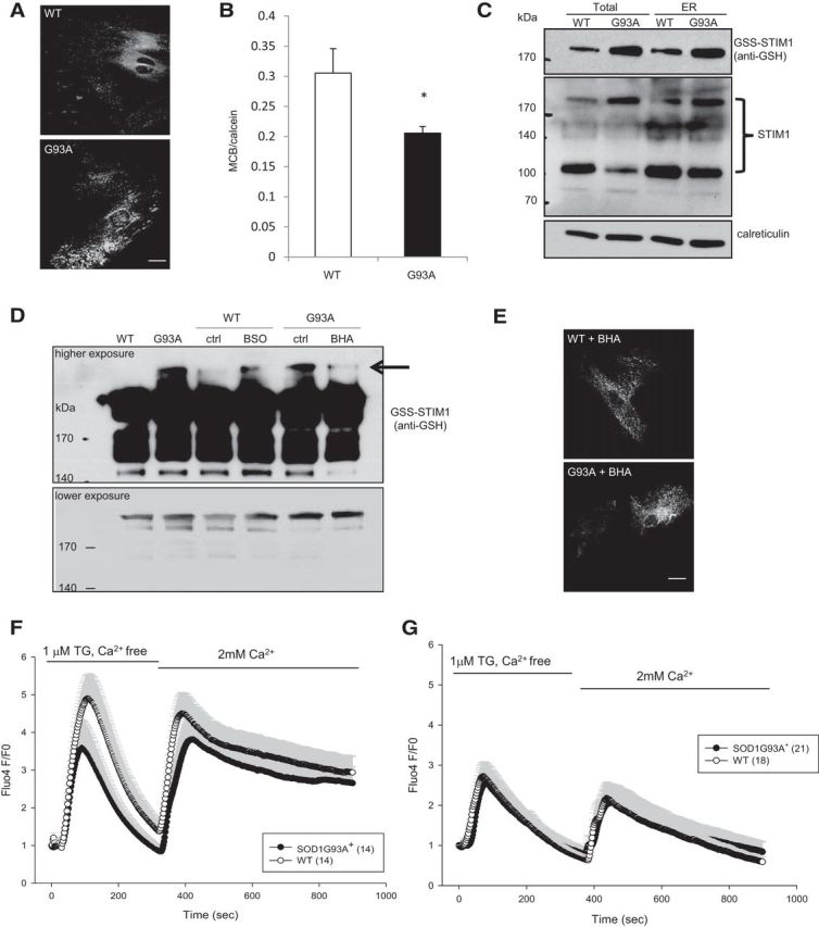
Glutathionylated STIM1 underlies abnormal SOCE in SOD1G93A+ astrocytes. A, WT and SOD1G93A+ astrocytes were transfected with YFP–STIM1 and fixed 48 h later. STIM1 localization was assessed under unstimulated conditions. Large clusters of YFP–STIM1 were observed in SOD1G93A+ astrocytes. B, Glutathione levels in spinal cord astrocytes determined by MCB and normalized to calcein fluorescence. C, ER/microsomes were enriched by fractionation from spinal cords of 60-d-old SOD1G93A+ and WT mice. Equal amounts of proteins from the postnuclear supernatant (TOTAL) and ER/microsome (ER) fractions were analyzed by nonreducing SDS-PAGE and Western blot. The middle panel shows multiple bands detected by the STIM1 antibody, including a monomeric species ∼100 kDa and higher-molecular-weight oligomers ∼200 kDa. The blots were reprobed with the anti-GSH antibody that confirmed the identity of the oligomeric STIM1 to be glutathionylated (top). Calreticulin was used as an ER marker (bottom). D, P2 cortical astrocytes were treated with 100 μm BSO (for WT cells) and 100 μm BHA (for mutant cells) for 18 h. Whole-cell homogenates were prepared in the presence of 50 mm NEM and used for immunoprecipitation with monoclonal anti-STIM1 antibodies. Immunoprecipitation samples were analyzed by nonreducing SDS-PAGE/Western blot using the anti-GSH antibody. Many forms of glutathionylated STIM1 was observed. The top and bottom panels show a representative blot, at high and low exposures, respectively. Oxidant-induced high-molecular-weight STIM1 oligomers (arrow) were detected in SOD1G93A+ astrocytes but not in WT cells. The level of these oxidant-induced STIM1 oligomers can be modified by pro-oxidant (BSO) and anti-oxidant BHA in WT and mutant astrocytes, respectively. ctrl, Control.E, Representative imagesof WT and SOD1G93A+of WT and SOD1G93A+ astrocytes transfected with YFP–STIM1, treated with 100 μm BHA (18 h). Cells were fixed 48 h after transfection to assess YFP–STIM1 localization. F, Cells were treated with 100 μm BSO for 4 h before imaging. BSO increased accumulation of ER calcium in WT cells, and SOCE was enhanced to levels similar to mutant cells. G, SOCE in cells treated with cell-permeable GSH (GSHEE; 1 mm) for 4 h. Mutant astrocytes SOCE normalized to WT SOCE levels when treated with GSH. All error bars are SEM. *p < 0.05 by Student's t test.