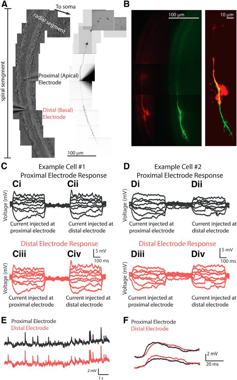
Figure 1.

Dual recordings from a type II afferent dendrite. A, Left, Tiled DIC image of the organ of Corti showing the placement of two recording electrodes, both in the spiral segment of dendrite, spaced ∼100 μm apart (average, 101.2 ± 7.6 μm). Right, Same field of view; fluorescent view of neuron. Alexa Fluor 488 hydrazide tracer signal is visible in the proximal electrode solution and filled type II dendrite. The second electrode contains Alexa Fluor 568 hydrazide and is not visible. B, Left, Both the Alexa Fluor 488 hydrazide (green) and the Alexa Fluor 568 hydrazide (red) tracers are visible in the same type II afferent dendrite, confirming a dual recording. A more complete fill is visible for the green tracer, from the electrode placed first. Right, Merged and enlarged image of green and red filled fibers (contrast enhanced). C, D, Passive flow of current in a single dendrite measured at two electrodes in two exemplar neurons. Ci, Cii, Voltage recording from the electrode proximal to the cell soma. Ci, Voltage response to current injected at this electrode. Cii, Voltage response to current injected at the other, distal electrode, 98 μm distant. Ciii, Civ, Voltage recording from the electrode distal to the cell soma. Ciii, Voltage response to current injection at the other electrode; passive propagation. Civ, Voltage response to current injection at this electrode. D, Same experimental design as in C for a different neuron (electrode spacing, 94 μm). E, Simultaneous measurement of EPSPs from two electrodes recording from the same dendrite. Top (black), Voltage recording from the proximal electrode showing EPSPs induced by local application of 40 mm KCl solution via a large-bore gravity-driven pipette. Bottom (red), Simultaneous voltage recording from the distal electrode. The patterns of EPSPs are the same at both electrodes. F, Exemplar EPSPs recorded simultaneously at the proximal (black) and distal (red) electrodes. Top, EPSP peaking first at the proximal electrode. Bottom, EPSP peaking first at the distal electrode.
