
Figure 1.
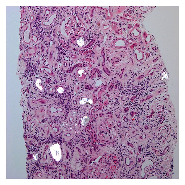
Hematoxylin and eosin stain of renal biopsy under polarized light (20x magnification) showing oxalate crystals in tubular lumen.
Official websites use .gov
A
.gov website belongs to an official
government organization in the United States.
Secure .gov websites use HTTPS
A lock (
) or https:// means you've safely
connected to the .gov website. Share sensitive
information only on official, secure websites.
Hematoxylin and eosin stain of renal biopsy under polarized light (20x magnification) showing oxalate crystals in tubular lumen.