Figure 2.

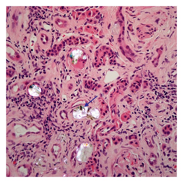
Hematoxylin and eosin stain of renal biopsy under polarized light (40x objective magnification) showing oxalate crystals with suggestion of “fan” like morphology (blue arrow).

Hematoxylin and eosin stain of renal biopsy under polarized light (40x objective magnification) showing oxalate crystals with suggestion of “fan” like morphology (blue arrow).