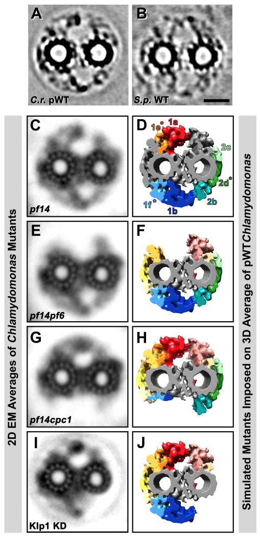
Fig. 7. Classical 2D EM averages of CPC mutants and simulation of corresponding 3D structural defects.
(A-J) Cross-sectional projection images through ~80 nm thick tomographic slices of CPC averages from Chlamydomonas pseudo-wildtype (C.r. pWT, A) and Strongylocentrotus wildtype (S.p. WT, B) are shown for comparison with 2D EM averages of ~80 nm thick plastic sections of the CPC from four Chlamydomonas mutants: pf14 (C) is a mutant lacking the radial spokes, but without CPC defects, i.e. the 2D averages looks like wildtype (compare with Fig. 1B). Note the similarities in densities amongst (A and C), as well as (B). The pf14pf6 (E), pf14cpc1 (G) double mutants and the Klp1 knockdown (I) all show CPC defects. Isosurface renderings of the Chlamydomonas pWT average have been edited by deleting selected projection densities (D, F, H and J) to reflect densities likely to be absent in the depicted Chlamydomonas CPC mutants (C, E, G, and I). The pf14pf6 double mutant in (E) appears to lack projections 1a and 1e. The 1b and 1f projections are almost completely absent in the pf14cpc1 double mutant (G and H). From the Kinesin-like protein 1 knockdown (Klp1 KD) (I) the 2b-d projections appear to be absent (J), which includes Hydin (see Fig. 8). Scale bar (B): 25 nm. Images from (C, E, and G) were adapted from [Mitchell and Sale, 1999] and image (I) was modified from [Yokoyama et al., 2004].

