
Figure 5.
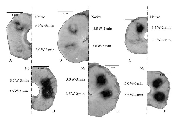
Dissection of formalin-fixed canine prostate tissue. Darkened areas indicate areas of thermal lesions induced at 3.0 or 3.5 W for 2 or 3 minutes. Sections A, B, and C indicate native tissue and sections D, E, and F indicate nanoparticle-laden tissue. 150 nm gold nanoshells were injected directly into the parenchyma of the prostate. It was found that lesion size grew with increasing power when applied to native tissue and that nanoshell application significantly increased the lesion size, but not with respect to power [23].