Figure 1.
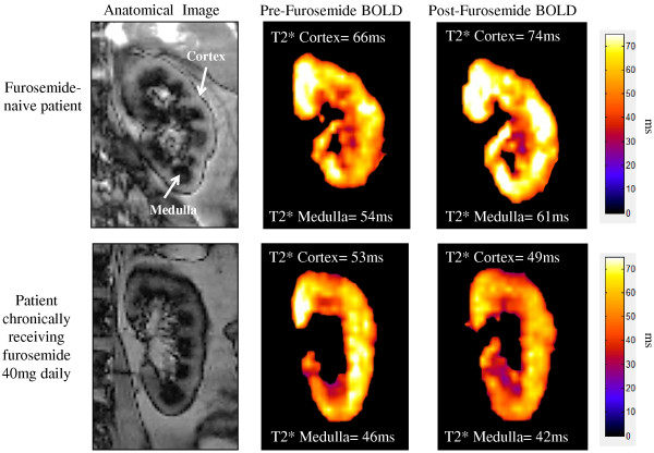
MR images of the left kidneys of two participants: furosemide naïve (top row) and chronic furosemide administration of 40 mg/day (bottom row). The anatomic images in the panel in the left column are generated from a non-contrast arterial spin labeling technique in which the renal cortical segments appear gray and the medullary segments appear dark (white arrows). Corresponding T2* maps representing tissue oxygenation (Blood Oxygen Level Dependent or BOLD) before (middle column) and after (right column) a 20 mg intravenous furosemide stimulus. On these images the color scale intensity represents the T2* value for that corresponding voxel. As shown, the furosemide naïve participant increased both cortical and medullary T2* signal intensities in response to a 20 mg intravenous furosemide stimulus by 12% and 13% respectively. However, the cortical and medullary T2* percent change in signal intensity decreased (bottom right panel) by 7% and 9% respectively after a 20 mg intravenous furosemide stimulus.

