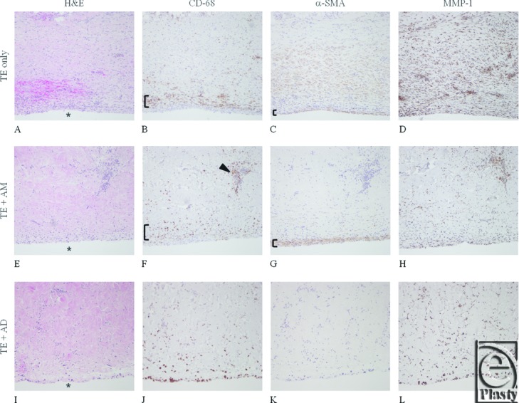
Figure 1.

Representative histology and immunohistochemical staining using serial sections of tissue overlying the implanted tissue expander (TE) interface (indicated by *) for the TE-only (A-D), AlloMax (AM; E-H), and AlloDerm (AD; I-L) groups. Tissues are stained/labeled for hematoxylin and eosin (A, E, I); CD68 epitope for macrophages (B, F, J); α-smooth muscle cell actin (SMA) for myofibroblasts (C, G, K); and matrix metalloproteinase-1 (MMP-1) for matrix collagenase (D, H, I). In the TE-only group, a rim of macrophages can be detected ([, panel B) and presence of myofibroblasts ([, panel C). Tissue around the AM-covered TE had more modest macrophage presence ([, panel F); however, clusters were observed (indicated by arrowhead in F) and myofibroblast staining was strong ([, panel G). The lowest signal for macrophages and myofibroblast was noted in tissues from AD-covered TE. Excessive MMP-1 staining (D, H, L) is suggestive of accelerated ECM degradation in TE-only and clustered activity in AM (not observed in tissue from AD-covered TE).
