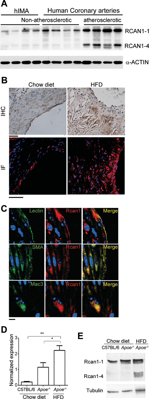
Figure 1.
RCAN1-4 is induced in atherosclerotic human and mouse arteries
Immunoblot of RCAN1 in atherosclerotic and non-atherosclerotic human coronary artery and in human internal mammary artery (hIMA). Protein bands corresponding to RCAN1-1 and RCAN1-4 are indicated. α-ACTIN was used as loading control.
The aortic sinus of Apoe−/− mice fed standard chow or a high-fat diet (HFD) was immunostained for Rcan1. Representative images are shown of Rcan1 immunohistochemistry (top panels) and fluorescence microscopy (bottom panels). Bar, 50 µm.
Representative confocal immunofluorescence images of Rcan1 (red), lectin (green), αSMA (green) and Mac3 (green) in aortic sinus lesions of HFD-fed Apoe−/− mice. Merged images are also shown. Bar, 5 µm.
Quantitative PCR analysis of Rcan1 mRNA expression in the aortic arch of C57BL/6 and Apoe−/− mice fed standard chow or an HFD. mRNA amounts were normalized to Hprt1 expression (means ± SEM; n = 3). Student's t-test, *p = 0.013, **p = 0.0019.
Representative Rcan1 immunoblot in the same tissues as in D.

