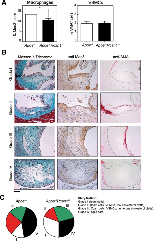
Figure 4.
Rcan1 deficiency results in less-advanced plaques
Quantification of neointimal macrophage and VSMC content in lesions of the aortic sinus of Apoe−/− (n = 11) and Apoe−/−Rcan1−/− mice (n = 18) fed an HFD for 6 weeks. Student's t-test, *p = 0.017.
Representative images of Masson's trichrome, anti-Mac3 and anti-SMA staining of lesions of the aortic sinus classified according to the Stary method: grade I (mostly foam cells), grade II (foam cells, VSMCs and few cholesterol clefts), grade III (foam cells, VSMCs and numerous cholesterol clefts) and grade IV (lipid core). Bars, 50 µm.
Classification of aortic sinus lesions according to the Stary method in the cohort of Apoe−/− and Apoe−/−Rcan1−/− animals shown in Fig 3.

