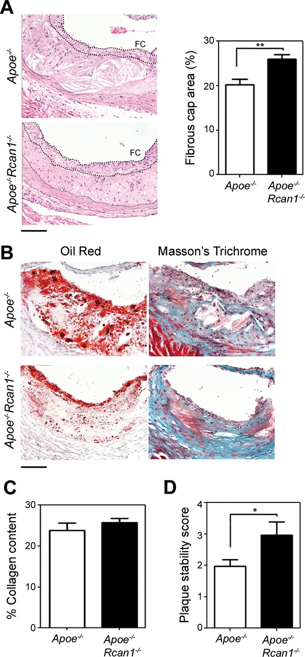
Figure 5.
Rcan1 deficiency does not decrease plaque stability
Representative H&E staining and quantification of fibrous cap area of lesions in the aortic sinus of Apoe−/− (n = 25 valves) and Apoe−/−Rcan1−/− mice (n = 45 valves) fed an HFD for 6 weeks. Data are shown relative to the lesion area (means ± SEM). Student's t-test, **p = 0.0012. Thick and thin dotted lines, respectively, delimit the fibrous cap (FC) and lesion.
Representative images of Oil Red and Masson's trichrome staining of cryocut cross-sections of the aortic arch lesions of Apoe−/− (n = 15) and Apoe−/−Rcan1−/− mice (n = 17) fed an HFD for 6 weeks. Bar, 50 µm.
Quantification of collagen content in the fibrous cap of these lesions (mean ± SEM).
Plaque stability score determined by collagen content divided by Oil Red staining area of the same lesions (mean ± SEM; Student t-test, *p = 0.048).

