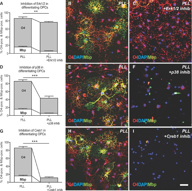
Figure 2.
Erk1/2, p38Mapk and Creb1 play a functional role during OPC differentiation.
Bar graph showing the decrease of Mbp-positive oligodendrocyte lineage (OL) cells after 2 days in differentiation medium in the presence of an Erk1/2 inhibitor. ANOVA: O4 p > 0.05, MBP **p < 0.001; Dunnett's post hoc test PLL versus PLL with ERK1/2 inhibitor: p < 0.0001; mean decrease: O4 = 11%; Mbp = 67%.
Bar graph demonstrating a significant decrease of O4/Mbp-positive OL cells after treatment with a p38 inhibitor. ANOVA: (O4 ***p < 0.0001, Mbp ***p < 0.0001; Dunnett's post hoc test PLL versus PLL with p38Mapk inhibitor: p < 0.0001; mean decrease: O4 = 49%, Mbp = 66%.
Bar graph indicating a significant decrease of O4 immunoreactivity and a complete loss of Mbp expression in the presence of a Creb inhibitor. ANOVA: O4 ***p < 0.0001, MBP ***p < 0.0001; Dunnett's post hoc test PLL versus PLL with Creb1 inhibitor: p < 0.0001; mean decrease: O4 = 87%, Mbp = 100%. Error bars: SEM.
Representative images of cells immunolabelled for O4 (red) and MBP (green) on control substrates and cultured in differentiation medium supplemented with ERK1/2 inhibitor. Scale bar = 30 µm.