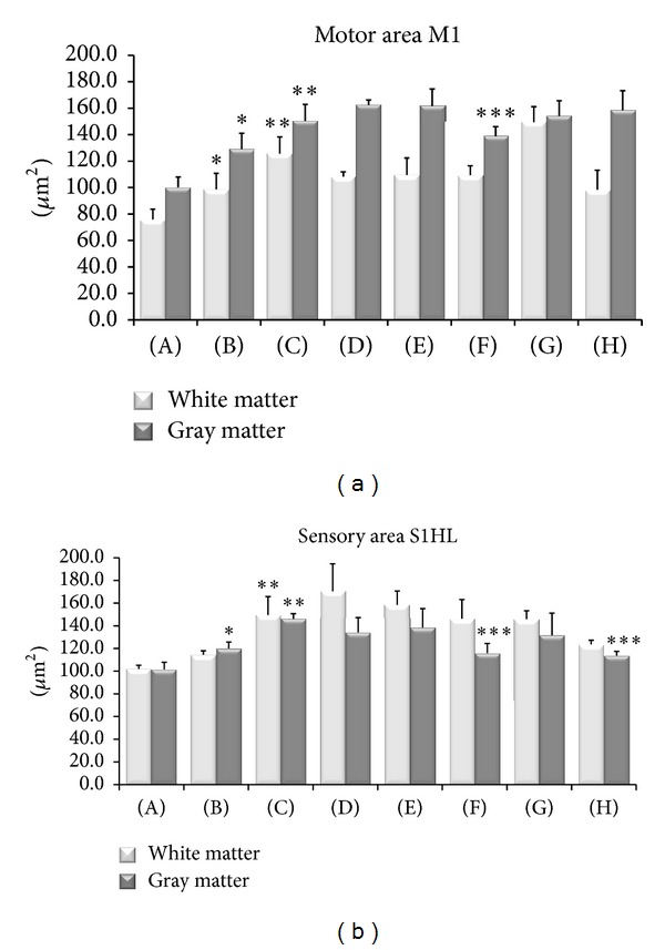
Figure 3.

Size of GFAP-immunoreactive astrocytes in the cerebral cortex primary motor and sensory areas. Mean of immunoreactions areas of astrocytes is expressed in μm2. Data are the mean ± SE. (A) Control Sham-operated WKY rats, (B) control Sham-operated SHRs, (C) control CCI SHRs, (D) CCI SHRs treated with (+/−)-thioctic acid 250 μmol/kg/day, (E) CCI SHRs treated with (+/−)-thioctic acid 125 μmol/kg/day, (F) CCI SHRs treated with (+)-thioctic acid 125 μmol/kg/day, (G) CCI SHRs treated with (−)-thioctic acid 125 μmol/kg/day, and (H) CCI SHRs treated with pregabalin 300 μmol/kg/day (H). The data of densitometric analysis were expressed as arbitrary units. *P < 0.05 versus WKY control rats; **P < 0.05 versus SHR Sham operated control rats; ***P < 0.05 versus control CCI SHR.
