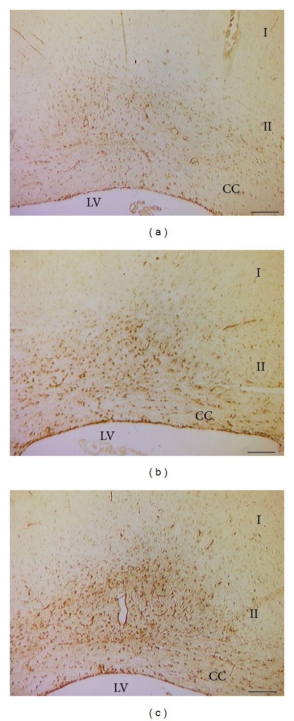
Figure 4.

Sections of the sensory cortex (S1HL) processed for the immunohistochemical demonstration of glial fibrillary acidic protein (GFAP). Note the increase of the immunoreactions in the zone II of the cortex in the control Sham-operated SHRs and in larger extent in the control CCI SHRs. (a) WKY control Sham-operated rats, (b) control Sham-operated SHRs, and (c) control CCI SHRs. I: zone 1; II: zone 2; CC: corpus callosum; LV: lateral ventricle. Calibration bar: 200 μm.
