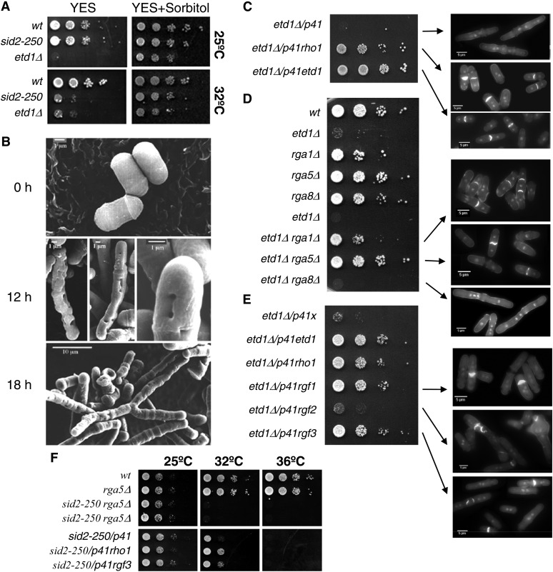
Figure 1.
Etd1-depleted cells are deficient for cell-wall integrity and septation, a phenotype suppressed by high Rho1 activity. (A) Growth assay (serial dilution drop tests on plates) of etd1Δcells in YES media (left panels) and YES with 1.2 M sorbitol (right panels). The wild-type and sid2-250 strains were used as controls. Cells were grown (at 37° for etd1Δ and wt and at 25° for sid2-250), and 10-fold dilutions (starting with 104 cells) were spotted and incubated at 25° or 32°. (B) Electron microscopy images of etd1Δ cells grown at 37° (permissive temperature for etd1Δ after 0-, 12-, and 18-hr incubations at 25° (restrictive temperature for etd1Δ). Etd1-deficient cells show severe defects in cell-wall integrity. (C) Growth assay (serial dilution drop tests on plates) of etd1Δ cells overexpressing Rho1 protein under nmt41x expression in the pREP41x vector (p41rho1, left). Cells were grown at 37°, and 10-fold dilutions (starting with 104 cells) were spotted and incubated at 30° in the absence of thiamine. The pREP41x-driven expression of Etd1 (p41etd1) and the empty pREP41x plasmid (p41) were used as positive and negative controls, respectively. DAPI- and Calcofluor-stained cells of these etd1Δ and etd1Δ/p41rho1 strains are shown (right panels). (D) Growth assay (serial dilution drop tests on plates) (left) and cell phenotype (DAPI and Calcofluor staining) (right panels) of etd1Δ rga1Δ, etd1Δ rga5Δ, and etd1Δ rga8Δ cells. Cells were grown at 37°, and 10-fold dilutions (starting with 104 cells) were spotted and incubated at 30°. Wild-type (wt) and single-deletion strains were used as control. (E) Growth assay (as described in C) and cell phenotype (DAPI and Calcofluor staining) (right panels of etd1Δ cells overexpressing Rgf1, Rgf2, or Rgf3 proteins under nmt41x expression in the pREP41x vector (p41rgf1, p41rgf2, and p41rgf3, respectivelly) (left). p41etd1 and p41rho1 were used as positive controls. The empty p41 plasmid was used as a negative control. (F) Growth assay of sid2-250 cells deleted for rga5 (single sid2-250 and rga5Δ mutant strains were used as controls) (top panels) or overexpressing rgf3 (p41rgf3) (bottom panels). The p41rho1 construct was used as a positive control. The empty p41 plasmid was used as a negative control. Cells were grown at 25°, and 10-fold dilutions (starting with 104 cells) were spotted and incubated at 25°, 32°, or 36° in the absence of thiamine in the case of overexpression assays (bottom panels).

