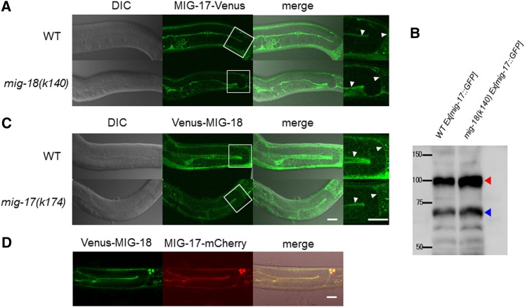
Figure 3.
Expression and localization of MIG-17-Venus and Venus-MIG-18. (A and C) DIC (left), confocal (center), and merged (right) images. The enlarged images of the boxed areas in the center panels are shown on the right side. The arrowheads point to the fluorescence from the gonadal basement membrane. (A) Wild-type and mig-18(k140) young adults that express MIG-17-Venus. (B) Western blot analysis of wild-type and mig-18(k140) animals that expressed MIG-17-GFP. Worm extracts (200 μg protein/sample) were analyzed using rabbit anti-GFP (2 mg/ml, Molecular Probes). The red and blue arrowheads indicate pro- and mature forms MIG-17-GFP, respectively. Intensities of the bands were quantified using ImageJ software. (C) Venus-MIG-18 expression in wild-type and mig-17(k174) animals. (D) Colocalization of Venus-MIG-18 and MIG-17-mCherry in the gonadal basement membrane. Confocal images for Venus (left) and mCherry (center) and DIC and confocal merged image (right). The strong dot-like signals on the upper right are of coelomocytes that take up secreted proteins. Posterior gonads are shown. Anterior to the left, dorsal to the top. Bar, 20 μm.

