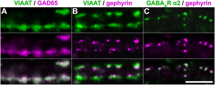
Figure 3. Convergence of immunofluorescence against VIAAT, GAD65, gephyrin, and the GABAA receptor α2 subunit on dendrites.

A-C, Representative confocal sections ∼1 um-thick showing the overlap of immunofluorescent puncta for VIAAT (green) and GAD65 (magenta) (A), VIAAT (green) and gephyrin (magenta) (B), and the GABAA receptor α2 subunit (green) and gephyrin (magenta) (C) on dendrites. Extensive overlap of puncta for VIAAT, GAD65, and gephyrin indicates that these antibodies label the same population of synapses in DIV20-22 dissociated hippocampal cultures. In addition, the overlap of puncta for GABAA receptor α2 and gephyrin suggests that GABAergic synapses were predominantly mature by this point. (Scale bar: 5 μm.)
