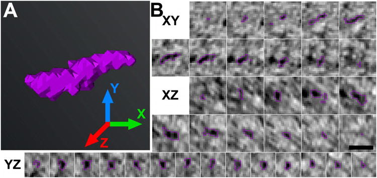
Figure 5. Segmentation in multiple orthogonal views enables accurate three-dimensional analysis of each postsynaptic filament.

A, Rendering of a discrete postsynaptic filament (violet) with X, Y, and Z axes indicated. B, Series of consecutive cross sections in the XY, XZ, and YZ views of the filament rendered in A. Cross sections were cropped from virtual sections 1.4 nm-thick, which were calculated from the tomographic reconstruction. The filament is outlined in violet for clarity. Segmentation in multiple planes permitted a more exact description of filaments' size, shape, and relation to other structures. (Scale bar: 20 nm.)
