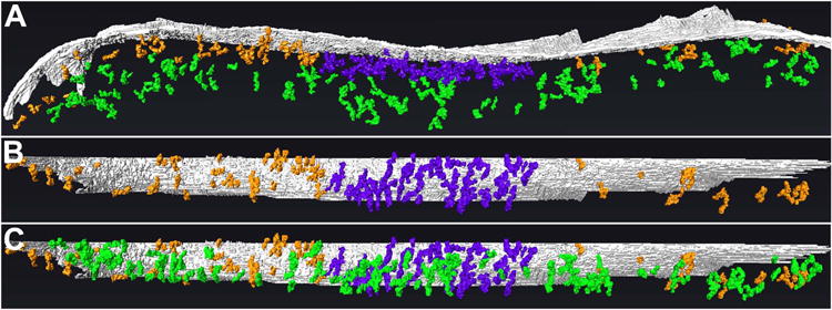
Figure 6. The postsynaptic network of filaments at GABAergic synapses consists of distinct zones.

A, Rendering (side-view) of the core, periphery, and mantle of the postsynaptic network. The core (rendered in violet), extending ∼30 nm into the cytoplasm from the postsynaptic membrane (gray), exhibits the greatest concentration of filaments and contacts between filaments. The periphery (orange), which appears to encircle the core, also extends ∼30 nm into the cytoplasm but exhibits a low concentration of filaments and inter-filament contacts. The mantle (green), located between ∼30 and ∼100 nm from the postsynaptic membrane and co- extensive with the core and periphery, exhibits fewer filaments and inter-filament connections than the core but more than the periphery. B, En-face view of postsynaptic membrane and network, omitting filaments of the mantle to visualize without obstruction the division between the core and periphery. C, En-face view of postsynaptic membrane and network, showing extent of the mantle.
