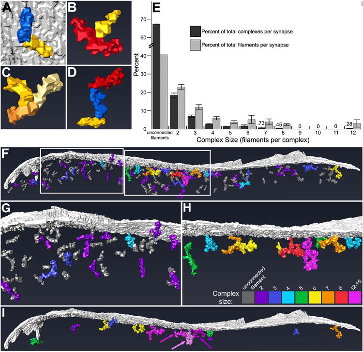
Figure 9. Filaments of the postsynaptic network at GABAergic synapses interconnect, forming higher-order complexes.

A, Rendering of short filament (in yellow) and medium filament (blue) contacting tip-to-tip. Opposite tips of both filaments also contacted the postsynaptic membrane (gray). B, Rendering of long filament (red) contacting a short filament (yellow) middle-to-tip. C, Rendering of three short filaments (yellow, light yellow, and orange) contacting tip-to-tip-to-tip, apparently trimerized. D, Rendering of short filament (yellow) and medium filament (blue) contacting tip-to-tip, with medium filament also contacting long filament (red) tip-to-tip. Such contacts allow filaments to assemble into what appear to be molecular complexes. E, Bar graph of percent of total complexes composed of different numbers of filaments (dark bars) and percent of total filaments incorporated into complexes of different sizes (light bars) (mean ± SEM). Unassociated filaments and complexes composed of ≤3 filaments outnumbered complexes composed of ≥4 filaments. The majority of filaments were either unconnected to others or incorporated into complexes made of ≤3 filaments. Values for the smallest data points are provided above the corresponding bars. F-H, Rendering (side view) of the postsynaptic network and membrane (gray) showing the distribution of mono- and poly- filament structures throughout (F). The number of filaments incorporated into a given complex is indicated by its color (see key in H). Complexes composed of ≥4 filaments (cyan-pink) were preferentially located in the core of the postsynaptic network, while those composed of ≤3 filaments (dark gray-blue) were more predominant in the periphery and mantle, but some were also intercalated between the larger complexes of the core. G and H depict the left and right boxed areas in F, respectively, in greater detail, with complexes composed of ≤3 filaments omitted for clarity in H. I, Rendering (side view) of the postsynaptic membrane (gray) and those complexes linked to others by irregular globules of electron-dense material; complexes that are not so linked have been excluded for clarity. The number of filaments incorporated into a given clutch of linked complexes (equal to the total number of filaments of its constituent complexes) is indicated by its color, according to the key in H. Arrows emphasize the distinction between three separate clutches rendered in pink, light pink, and dark pink.
