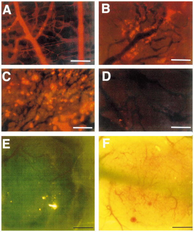
Fig. 7.
Transvascular transport in dorsal skin and tumors. (a) There is hardly any extravasation of 90-nm diameter liposomes from normal vessels; (b) heterogeneous extravasation of 90-nm diameter liposomes from LS174T tumor vessels, 48 h after injection. Note that some vessels are leaky as indicated by the yellow fluorescence for rhodamine, while others are not. Extravasated liposomes do not diffuse far from blood vessels (adapted from Ref. [14]). (c) Liposomes of 400 nm diameter (yellow fluorescent spots) extravasate adequately from LS174T tumor. (d) Liposomes of 600 nm diameter do not extravasate, suggesting that LS174T vessels have pore-size cut-off of about 500 nm (adapted from Ref. [66]). (e) The human glioma (HGL21) xenograft is permeable to Lissamin green (i.e., tumor tissue becomes green) when grown subcutaneously (Yuan and Jain, unpublished results). (f) The same glioma develops blood–brain barrier properties (i.e., impermeable to Lissamin green) when grown in the cranial window (adapted from Ref. [14]).

