Figure 1.
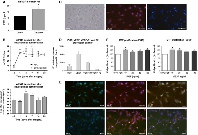
Placental growth factor (PlGF) plays an important role in scar formation in vitro. (A) PlGF levels were up-regulated in aqueous humour of glaucoma patients (*P = 0.03 versus control participants; n = 10 per group). (B) Bevacizumab treatment induced a significant increase in aqueous PlGF levels on day 3 (1.3-fold) and 7 (1.2-fold), as compared with control eyes injected with NaCl (*P < 0.05 versus control; n = 5). (C) The cultures of primary murine Tenon fibroblasts (MTF) clearly show an adherent homogeneous morphology of spindly, generally flat, elongated shaped cells (left panel). The cells are immunopositive for vimentin (red; middle panel), but do not show any staining for the epithelial cell marker, cytokeratin (red; right panel). Scale bar: 50 μm. (D) VEGF, PlGF and their receptors (VEGF-R1 and -R2) were expressed by MTF. The mRNA levels were normalized to that of the house keeping gene β-actin. (E) Tenon fibroblasts were immunopositive for vimentin (red; middle panels) and VEGF and PlGF (green; left panels). VEGF and PlGF both colocalized with vimentin in the cytoplasm of the cells (merge; right panels). 4′,6-diamidino-2-phenylindole (blue) was used a nuclear counter staining. Scale bar: 50 μm. (F) Addition of recombinant murine VEGF and PlGF (10–100 ng/ml) significantly augmented MTF proliferation [*P < 0.05 versus the control medium, containing 0.1% FBS (white bar)].

