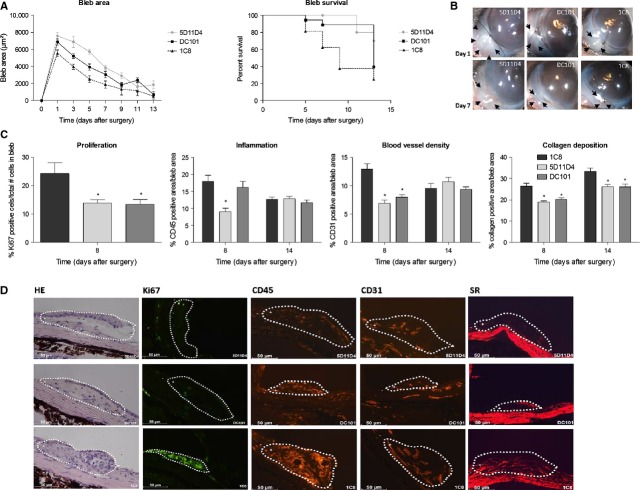
Figure 4.
Single intracameral injection of anti-PlGF antibody improves surgical outcome in a mouse trabeculectomy model. (A) Bleb area was significantly larger at each time-point compared with control after administration of the anti-PlGF antibody (P < 0.001; n = 20), whereas anti-VEGF-R2 antibody did not significantly improve bleb area (P = NS; n = 20). 5D11D4 also significantly prolonged bleb survival (P = 0.002; n = 20), while DC101 did not (P = NS; n = 20). (B) It shows macroscopic post-operative photographs of the blebs on days 1 and 7 after surgery. Arrows: edges of the blebs. (C) Treatment of 5D11D4 significantly decreased the process of inflammation at the filtration site on day 8, whereas DC101 did not. Proliferation, blood vessel density and collagen deposition were reduced after single administration of anti-PlGF and anti-VEGF-R2 antibody (*P < 0.05 compared to 1C8; n = 10 per time point). (D) The images show representative pictures of immunostainings of eyes treated with 5D11D4 (upper panels) or with DC101 (middle panels) or control eyes (lower panels), at 8 days after surgery. Edges of the blebs are marked by a dotted line and were indicated as the positive area of analysis (+), whereas the rest of the eye was indicated as the negative area of analysis (−). Scale bar: 50 μm.

