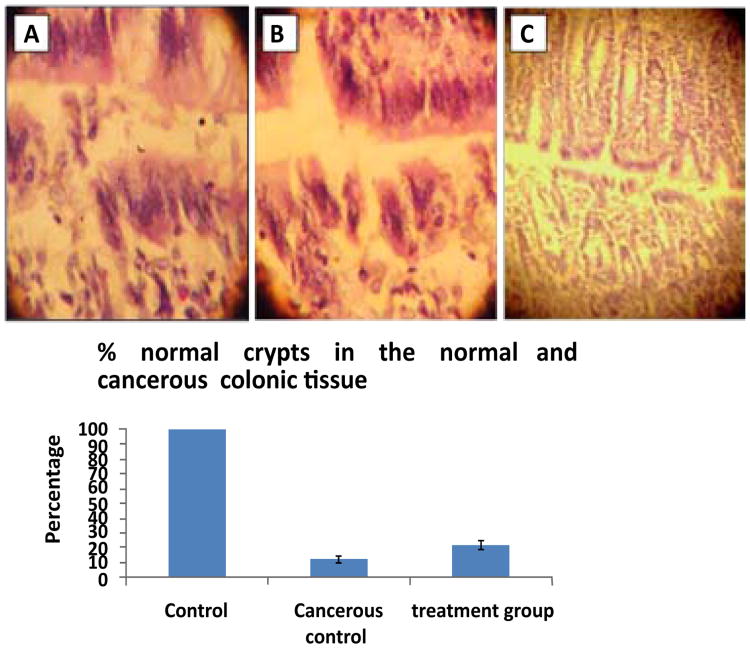
Figure 2. Histopathology of colon cancer induced in rats.
Panel (A & B). Treated groups of cancerous colon showing aberrant crypt foci formation. Panel (C). Normal colon cancer showing aberrant crypt foci formation after the treatment (that were shown as normal pathology). This is the representative histopathology from a field of 100× magnification. All animals in the respective group showed similar histopathology with no variations and experiments were repeated three times.

