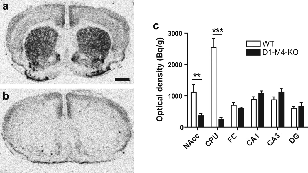
Fig. 6.
In situ hybridisation autoradiograms showing M4 mRNA levels in treatment-naive floxed WT (a) and D1-M4-KO (b) mice at the level of the dorsal striatum. Magnification bar = 1 mm. (c) Quantification revealed a prominent reduction in the nucleus accumbens (NAcc) and the caudate-putamen (CPU) in D1-M4-KO mice compared to WT mice while there were no significant differences in the frontal cortex (FC), hippocampal dentate gyrus (DG) and subfields CA3/CA1. Data represent group mean ± SEM. **p<0.01, ***p<0.001 vs. WT (Student’s t-test; n=6)

