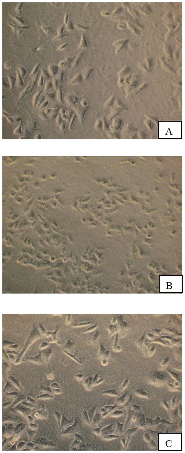
Figure 3. Inverted microscopy reveals morphological changes on HEp-2 cells after infection with WT (B) and VCUSM21P (C), at a magnification of ×40.

As control, HEp-2 cells in PBS (A) were included in the cytotoxic assay. HEp-2 cells were incubated for 1 hour with WT and VCUSM21P cells at multiplicity of infection (MOI) of 100. B shows rounded HEp-2 cells indicating cytotoxic effects. A and C show HEp-2 cells with normal morphology indicating absence of cytotoxicity.
