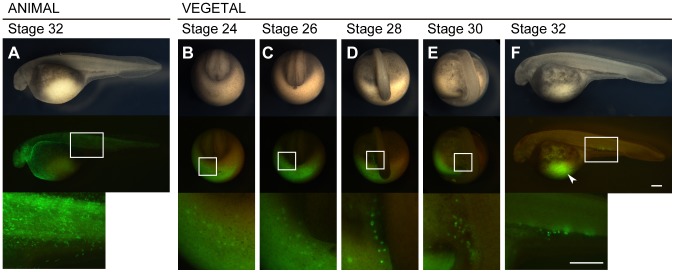
Figure 2. Migration of visualized PGCs in sturgeon embryo.
An embryo injected with the GFP-nos3 3′UTR mRNA at the animal pole (A) and at the vegetal pole (B–F). (A) The animal pole labeled embryo at stage 32. (B) The vegetal pole labeled embryo at stage 24. PGCs were initially found around the marginal region of the posterior developing embryonic body at this stage (C) The embryo at stage 26. The PGCs migrated dorsally as the tail rudiment bulged out. Until this stage, the distribution of the labeled PGCs is crescent-like surrounding the developing tail bud. (D) The embryo at stage 28. The PGCs were divided into two populations, at the left- and right-side of the embryonic body. At this stage, fluorescence of PGCs was stronger than during stages 24 and 26. Most PGCs are still localized on the yolk ball. (E) The embryo at stage 30. Most PGCs are located on the yolk extension, but some more ventral were still on the yolk ball. (F) The embryo at stage 32. PGCs are localized at the position where the gonads will develop. Some of these cells migrated axially. Note that PGCs migrated a long distance from their position of origin (arrowhead). The upper, middle, and lower columns indicate bright views, fluorescent views, and magnified fluorescent views of the boxes in the middle column, respectively. B-E are posterior views. E and F are lateral views. The scale bars indicate 500 µm.

