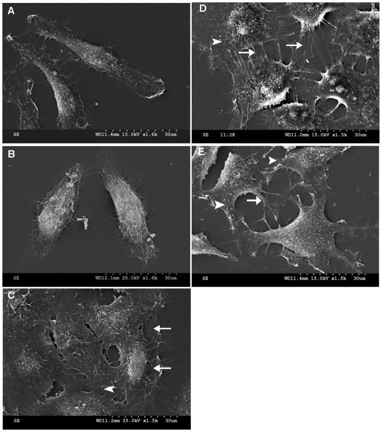
Figure 1. MF induces increases in filopodia-like protrusions and focal adhesions inFL cells.
A: negative control (N-con); B: sham-exposed (Sham); C: treated with100 nMEGF (EGF); D: exposed to 0.4 mT MF for 30 min (MF); E: pre-treated with PD, then exposed to MF (+PD+ MF). Arrow: appearance of filopodia; arrowhead: lamellipodia. The detailed information of experimental conditions and repeating numbers of samples is seen in Table 1.

