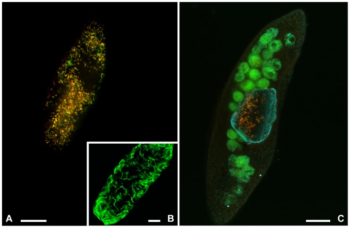
Figure 3. FISH experiments performed on “Candidatus Trichorickettsia mobilis” and “Candidatus Gigarickettsia flagellata”.
Symbionts inside P. nephridiatum PAR (A) and inside P. multimicronucleatum LSA (C) with probe RickFla_430 (Cy3, red signal) together with eubacterial probe EUB338 (fluoresceine, green signal). Food bacteria, labeled only in green, are visible inside food vacuoles in LSA (C). FISH experiment performed on “Candidatus Gigarickettsia flagellata” with probe GigaRick_436 (green signal) is shown in B. Bars: 20 µm.

