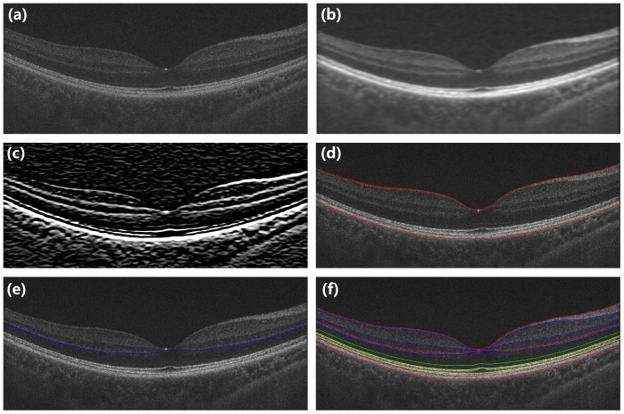
Figure 2. The detailed sequence in the boundary segmentation process.
(a) Original image. (b) Image smoothing. (c) Gradient image. (d) The ILM and the boundary between the RPE and choroid layers were first segmented. (e) Limiting detection area and search the minimum-weighted path. (f) Segmented image.

