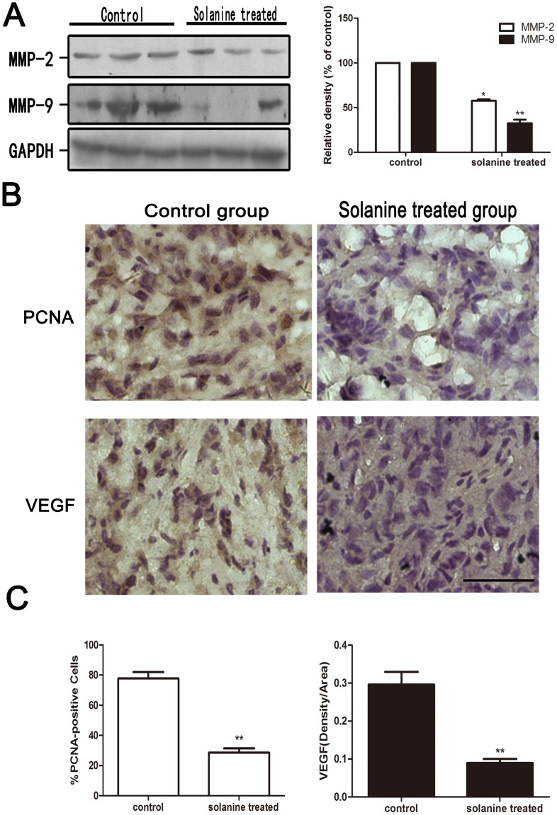
Figure 9. Effect of α-solanine on the expression of MMP-2, MMP-9, PCNA and VEGF in PANC-1 tumor xenograft.
When tumor xenografts were seperated, parts of tumor were used for Western blot and Immunohistochemistry. (A) The expressions of MMP-2 and MMP-9 protein were analyzede by Western blot. (B,C) Tumor tissus were innunohistochemically analyzed for PCNA-positive cells and VEGF staining density. Representative photographs of IHC staining of PCNA and VEGF are shown at 400×magnifications. Scale bar: 50 µm. The data are presented as the mean ± SE. *p<0.05, **p<0.01, compared with the control.

