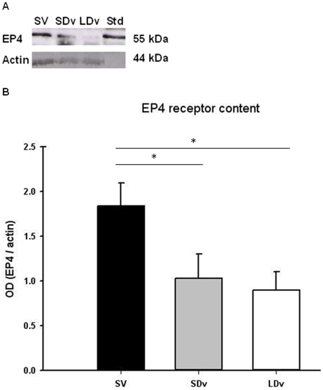
Figure 2. EP4 receptor decreased in varicose veins.
Protein measurements, representative samples of Western blot (A) of EP4 receptor in human small and large diameter varicosities (paired SDv and LDv, n = 7) and healthy saphenous veins (SV, n = 4). Western blot quantification of EP4 (B). Optical density (OD, arbitrary units) was quantified by Scion Image® and normalized by α-actin; * P<0.05 as determined by one-way-ANOVA followed by the Tukey post-hoc test and by a paired t-test for varicose veins.

