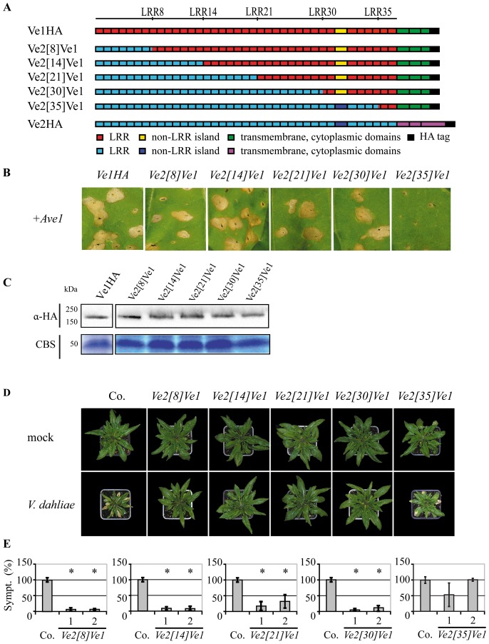
Figure 4. Functional characterization of Ve chimeric proteins that contain the C-terminus of Ve1.
(A) Schematic representations of transgenically expressed Ve1 (Ve1HA) and Ve2 (Ve2HA) and the proteins encoded by the chimeric genes Ve2 [8] Ve1, Ve2 [14] Ve1, Ve2 [21] Ve1, Ve2 [30] Ve1, and Ve2 [35] Ve1. The numbers indicate the eLRR at the site of the swap. (B) Typical appearance of tobacco leaves coinfiltrated with chimeric genes and Ave1. Pictures were taken at five days post infiltration, and show representative leaves for least 3 independent infiltrations. (C) Stability of chimeric Ve proteins is shown by immunoblotting using HA antibody (α-HA). Coomassie-stained blots (CBS) showing the 50 kDa Rubisco band present in the input samples confirm equal loading. (D) Typical appearance of non-transgenic sgs2 (Co.) and transgenic Arabidopsis sgs2 lines upon mock-inoculation or inoculation with V. dahliae race 1. Photographs were taken at three weeks post inoculation and show a representative plant of the non-transgenic sgs2 as well as a representative plant from one of the independent transgenic lines. (E) Quantification of Verticillium wilt symptoms (Sympt.) in Co. and transgenic lines. Bars represent quantification of symptoms presented as percentage of diseased rosette leaves with standard deviation. Co. is set to 100%. Asterisks indicate significant differences when compared with Co. (P<0.001). For each construct two independent transgenic lines are shown (1, 2).

