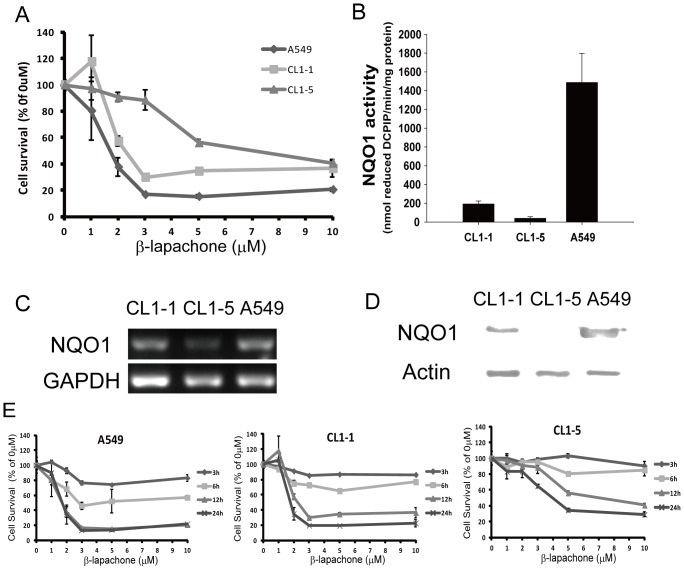
Figure 1. β-lapachone-induced cell death is associated with NQO1 expression levels.
(A) Percentage survival of the lung cancer cell lines CL1-1, CL1-5, and A549. Cells were treated with 0–10 µM β-lapachone for 12 h, then cell viability was determined by crystal violet staining assay and expressed as a percentage of the value for cultures with no β-lapachone. (B–D) NQO1 activity levels (B), NQO1 RNA expression levels (C), and NQO1 protein expression levels (D) in the three lung cancer cell lines grown under normal culture conditions. (E) Percentage survival of A549 cells (left panel), CL1-1 cells (center panel), and CL1-5 cells (right panel) incubated with the indicated concentration of β-lapachone for 3, 6, 12, or 24 h examined by crystal violet staining and expressed as percentage survival compared to the untreated cells. The results are the mean ± SD for 3 independent experiments, each in triplicate.

